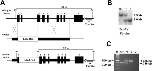
Figure 1.
Targeted disruption of the mouse Best2 gene. (A) The wild-type locus, targeting vector, and mutant locus. Thick lines: fragments used for constructing the targeting vector 5′ and 3′ arms. Thin lines: genomic DNA or vector backbone sequence. Numbered solid boxes: Best2 exons. Labeled boxes: the LacZ and neor expression cassette. The external 3′ probe used for Southern blot analysis in (B) is indicated under the wild-type and mutant loci. Filled arrows at exons 3 and 4 of the wild-type locus and LacZ-neor and exon 4 of mutant locus indicate the PCR primers used for genotyping in (C). Open arrows at exons 1 and 5 of wild-type locus indicate the primers used for RT-PCR in Figure 2. RV, EcoRV. (B) Southern Blot analysis was performed to identify homologous recombinants. Mouse genomic DNA was digested with EcoRV. Wild-type (+/+) fragment, 8.9 kb; mutant (−/−) fragment, 7.0 kb. The heterozygous mice (+/−) had both fragments. (C) Genotyping was confirmed using a multiplex PCR with two forward primers in exon 3 and the LacZ and neor expression cassette, respectively, and one reverse primer in exon 4. The 386-bp product represents the wild-type allele and is absent from the Best2−/− mice. The 486-bp product representing the disrupted allele is present only in the Best2+/− and Best2−/− mice. M, molecular weight marker.

