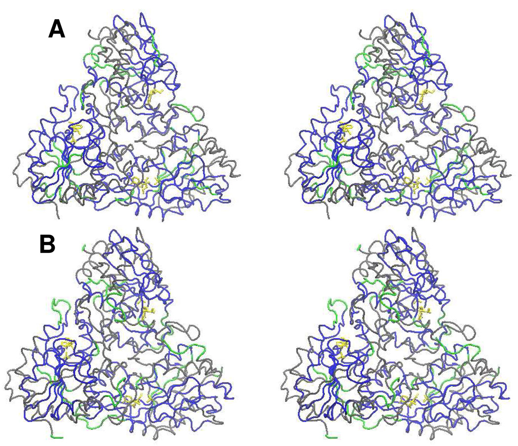
FIGURE 4.
Stereo tube diagram of the three-dimensional structure of the human PNP trimer in the presence of ImmH and phosphate (Panel A) or in the presence of DATMe-ImmH and phosphate (Panel B). ImmH and phosphate are shown in yellow in all catalytic sites in Panel A. Exchange sites based on H/D exchange properties are color-coded from the peptide H/D exchange data. Grey represents peptides with no change in deuterium uptake as a consequence of Immucillin binding. Blue represents peptides strongly protected from H/D exchange as a consequence of Immucillin binding. Green peptides indicate no sequence coverage. Most exchange occurs at the surfaces exposed to solvent and most reductions in exchange occur at the catalytic/binding sites. In B, DATMe-ImmH and phosphate are shown in yellow in all catalytic sites. Exchange sites based on H/D exchange properties are color-coded as above from the peptide H/D exchange data. Most exchange in the presence of DATMe-ImmH and phosphate occurs at the surfaces exposed to solvent and most reductions in exchange occurs at the catalytic/binding sites.

