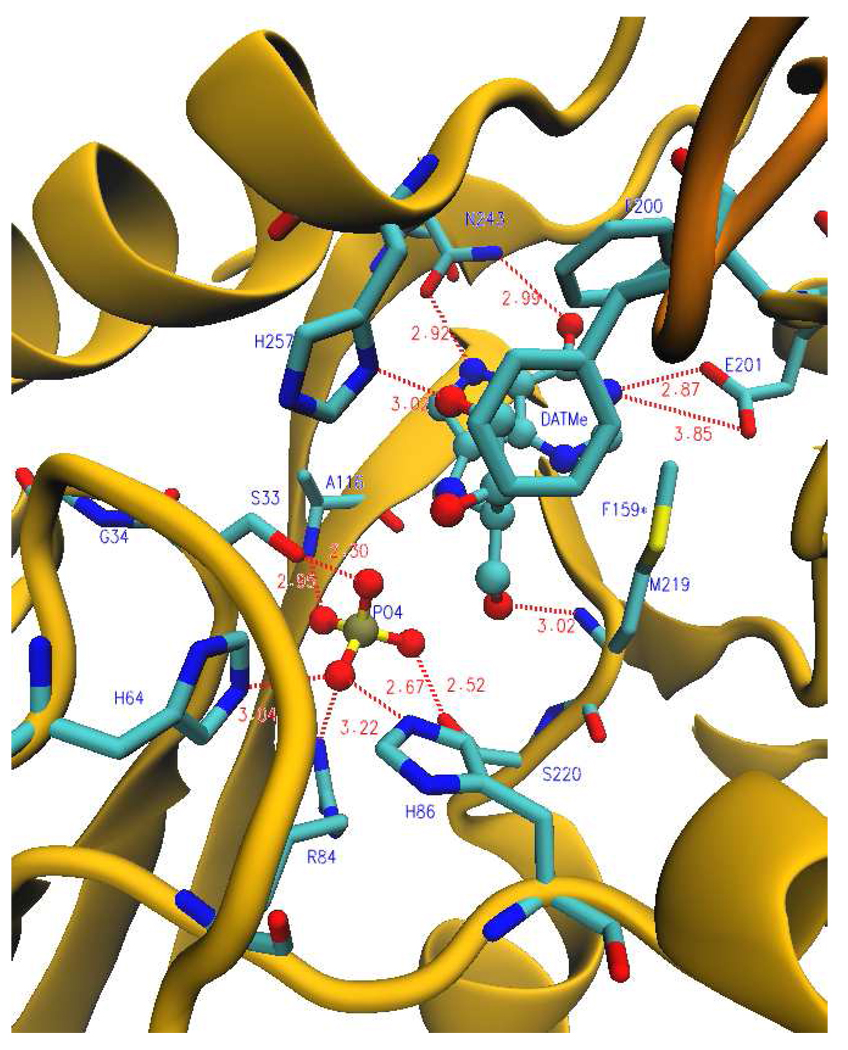
FIGURE 7.
Ribbon diagram showing a catalytic site of human PNP in the presence of DATMe-ImmH and phosphate. Blue labels (residues protected during H/D exchange); dark yellow (ribbon structure of monomer); orange (ribbon structure of neighboring monomer); cyan (carbon backbone); blue (nitrogen atoms); red (oxygen atoms); yellow (sulfur atom); gold (phosphorus atom). F159* is protected from the neighboring subunit. Dashed lines indicate hydrogen bonds or ionic interactions and distances are shown in angstroms (from PDB code: 2oyd). In the DATMe-ImmH complex loop 50–65 is in its closed form bringing H64 and G34 in catalytic contact to be protected (Figure 10). These residues are not protected in the ImmH complex (Figure 8).

