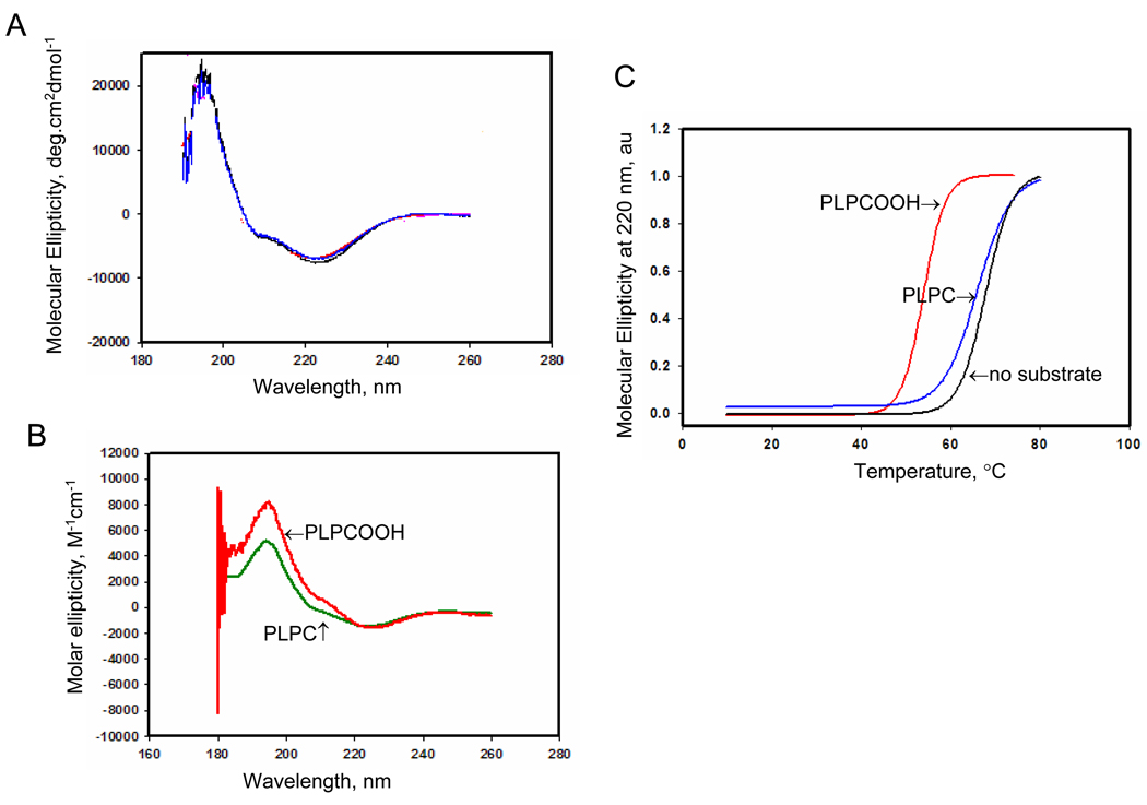
Figure 2. Circular dichroism (CD) analysis of Prdx6 in the presence of substrate.
A. CD spectrum of Prdx6 (1.25 mg/ml) in 40 mM phosphate buffered saline, pH 7.4. Spectra are shown for wild type (WT) (blue) and C47S mutant (red) proteins. The spectra are essentially identical. B. CD spectrum for wild type Prdx6 in the presence of reduced (PLPC) or oxidized (PLPCOOH) substrate (200 µM). C. Melting temperature for wild type protein derived from the spectra shown in A and B. Each melting curve was normalized to its maximal CD signal and the melting point was determined as the temperature at 50% loss of molecular ellipiticity (indicated by the arrows). The Prdx6 used in these assays was enzymatically inactive. All results are representative of 3 independent experiments.

