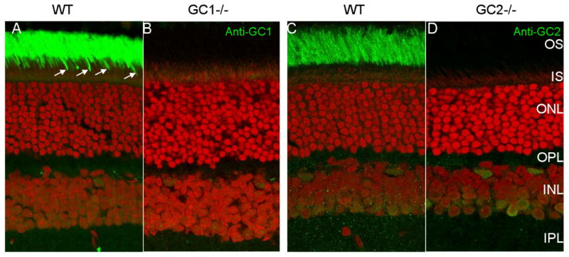
Figure 2.

Distribution of GC1 and GC2 in WT (A,C) and GC knockout (B,D) mouse retina. Cryosections A,B were probed with anti-GC1, and C,D with anti-GC2 antibodies. GC1 is present in rod (intense green staining in the OS area) and cone (arrows) outer segments (A). GC2 is only detectable in WT rod outer segments (C). GC1 and GC2 are undetectable in the ONL or OPL (synaptic terminals). Faint immunofluorescence in the INL is nonspecific.
