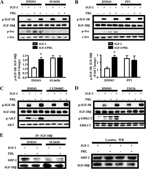
FIGURE 4.
SFKs mediate PRL enhancement of IGF-IR phosphorylation and SHP-2 association with IGF-IR. A–D, serum-starved MCF-7 cells were pretreated with DMSO and either 10 μm SU6656 (A), 10 μm PP1 (B), 10 μm U0126 (C), or 10 μm LY294002 (D) for 1 h prior to treatment with vehicle, IGF-I, PRL, or IGF-I/PRL for 15 min. The immunoblots were performed using cell lysates and the indicated antibodies (representative experiments shown). p-IGF-IR signals induced by IGF-I/PRL co-treatment were quantified and compared with those induced by IGF-I alone as described under “Experimental Procedures” (means ± S.D., n = 5 (A) or n = 3 (B)). The asterisks denote significant differences compared with IGF-I treatment. *, p < 0.05; **, p < 0.01) two-way ANOVA, Bonferroni post-test). E, serum-starved MCF-7 cells were pretreated with dimethyl sulfoxide (DMSO) or 10 μm SU6656 for 1 h prior to hormone treatment for 15 min. 1 mg of protein from cell lysates was immunoprecipitated (IP) with 1 μg of IGF-IRβ antibody and subjected to immunoblotting as indicated. Top panel, immunoblots of representative immunoprecipitation results; bottom panel, preimmunoprecipitation lysates. WB, Western blot.

