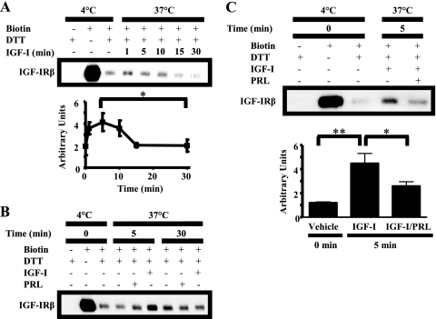
FIGURE 6.
PRL decreases IGF-I-induced IGF-IR internalization. Serum-starved MCF-7 cells were labeled with 0.25 mg/ml sulfo-NHS-biotin at 4 °C for 2 h. The cells were then treated for the times indicated with vehicle (B), IGF-I (A–C), PRL (B), or IGF-I/PRL (C) at 37 °C to allow for internalization of IGF-IR. Remaining plasma membrane-associated biotin was then cleaved with dithiothreitol (DTT). Biotinylated proteins that had internalized were immunoprecipitated using NeutrAvidin-conjugated beads and subjected to Western analysis (representative experiments shown). Internalized biotinylated IGF-IR was quantified using densitometry. A, means ± S.D. (n = 3). The asterisk denotes a significant difference in internalized biotinylated IGF-IR between 5 and 30 min. *, p < 0.05 (paired t test). C, means ± S.D. (n = 3). The asterisks denote significant differences in internalized biotinylated IGF-IR compared with IGF-I treatment. *, p < 0.05; **, p < 0.01 (one-way ANOVA, Neuman-Keuls post-test).

