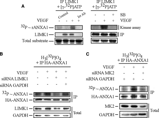
FIGURE 4.
Immunoprecipitated LIMK1, activated in response to VEGF, phosphorylates annexin 1 downstream of MAPKAP kinase-2. A, quiescent HUVECs were pretreated or not pretreated for 1 h with the p38 inhibitor SB203580 (SB, 1 μm). HUVECs were then treated or not treated with VEGF (5 ng/ml for 15 min). After treatments, cells were lysed, and LIMK1 was immunoprecipitated (IP) using rabbit polyclonal antibody and was subjected to an in vitro kinase assay. Reaction mixtures for kinase assays were put in the presence of [γ-32P]ATP, and LIMK1 activity was determined in immunocomplex assays using 5 μg rANXA1. Protein mixtures were separated through SDS-PAGE and were transferred to a nitrocellulose membrane. Kinase activity was then quantified by autoradiography using the PhosphorImager system by measuring [32P] incorporation into rANXA1. Membrane was also processed for immunodetection of immunoprecipitated LIMK1. Total rANXA1 is shown as an internal control for the amount of added substrate. A control track containing all kinase assay compounds except immunoprecipitated kinase is shown (Control). Immunoprecipitation using a rabbit isotype irrelevant antibody (Irr Ab) shows the specificity of the anti-LIMK1 antibody. In vitro phosphorylation of ANXA1 by LIMK1 was obtained in five distinct experiments. Representative results of those experiments are shown. In B, HUVECs were co-electroporated with siRNAs that target human GAPDH or LIMK1 mRNAs (200 pmol of siRNA) together with pIRES-hrGFP-2a containing HA-tagged wild-type ANXA1 construct. In C, HUVECs were co-electroporated with siRNAs that target human GAPDH or MAPKAP kinase-2 (MK2) mRNAs (80 pmol of siRNA) along with pIRES-hrGFP-2a containing HA-tagged wild-type ANXA1 construct. After 48 h, in B and C, cells were serum-starved for 16–20 h before being assayed for in vivo phosphorylation, as described under “Experimental Procedures.” Cells were treated or not treated with VEGF (5 ng/ml for 15 min). Thereafter, proteins were extracted, and the HA-tagged proteins were immunoprecipitated and processed as described in Fig. 2B. Incorporation of [32P] into HA-ANXA1 (upper panel) and immunoprecipitated HA-ANXA1 (second panel) are shown. The efficiency of the siRNA knockdown of LIMK1 and GAPDH (B) or MAPKAP kinase-2 and GAPDH (C) was determined (lower two panels) in Western blotting as described above using the total cell extracts. Representative results of at least three separate experiments are shown in B and C. MK2, MAPKAP kinase-2.

