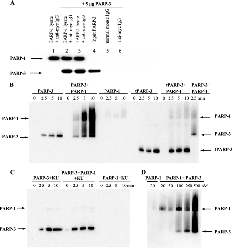
FIGURE 3.
Interaction between PARP-3 and PARP-1. A, shown is the immunoprecipitation of PARP-3 with PARP-1. Myc-PARP-1 was pulled out from U2OS cell lysate by anti-Myc antibody-agarose beads, and purified PARP-3 was added. The protein interaction was analyzed by Western blotting. Lane 1, no PARP-3 added; lanes 2 and 3, PARP-3 incubated with Myc-PARP-1 and anti-Myc antibody beads and washed with 1 m NaCl (lane 2) or 0.1% SDS (lane 3), respectively; lane 4, PARP-3 only; lanes 5 and 6, PARP-3 incubated with normal IgG-agarose beads or anti-Myc antibody-agarose beads, respectively. B, shown are the results from the activity assay using PARP-3, tPARP-3, and PARP-1 in the absence of activated DNA. PARP-3 or tPARP-3 was incubated with Bio-NAD+ and NAD+ in the absence or presence of PARP-1. At time 0 and after 2.5, 5, and 10 min of incubation at room temperature, the reaction was stopped by dilution in LDS sample buffer. C, to selectively inhibit PARP-1 activity, the protein was preincubated with 50 nm KU0058948 (KU) before the activity assay was performed. D, the activity of 20 nm PARP-1 was assayed for 5 min in the presence of different concentrations of PARP-3.

