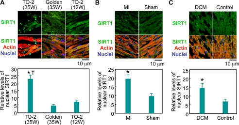
FIGURE 1.
Nuclear expression of SIRT1 in failing hearts. Left ventricular sections were stained with an anti-SIRT1 antibody (green), phalloidin-TRITC (actin, red), and Hoechst 33342 (nucleus, blue). A, representative immunostainings of 35-week-old TO-2 hamsters suffering from severe heart failure, control golden hamsters, and 12-week-old TO-2 hamsters that had not yet developed heart failure. B, non-necrotic, viable areas of rat hearts were analyzed 4 weeks after myocardial infarction (MI) or a sham operation (Sham). C, immunostaining of left ventricular muscles from a DCM patient and a patient without heart disease. In the bottom panels, quantitative analyses of the level of nuclear SIRT1 relative to that of cytoplasmic SIRT1 are shown. *, p < 0.05 versus age-matched (35-week-old) golden hamsters, sham-operated rats, or control patient. †, p < 0.05 versus young (12-week-old) TO-2 hamsters that had not yet developed heart failure; W, weeks.

