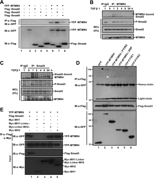
FIGURE 3.
MTMR4 interacts with Smad2/3. A, MTMR4 interacts strongly with Smad2/3. HEK293T cells transiently expressed Flag-tagged Smad2 (0.8 μg), Smad3 (0.8 μg), or Smad4 (0.8 μg) and YFP-MTMR4 (1.5 μg). Whole cell lysates were immunoprecipitated with M2 Flag antibody, and resolved proteins were immunoblotted with anti-GFP antibody (top panel). Protein expression levels were verified by anti-GFP (YFP-MTMR4, middle) or -Flag (Smads, bottom) antibodies, respectively. B and C, endogenous MTMR4 interacts with endogenous Smad2/3 upon TGF-β1 stimulation. Cell lysates were prepared from 90% confluent HaCaT cells after TGF-β1 treatment (2 ng/ml) for different lengths of time as indicated. Endogenous Smad2/3 bound to endogenous MTMR4 was immunoprecipitated with anti-MTMR4 antibody (B) or anti-Smad3 antibody (C) and detected by Western blotting with anti-Smad2 antibody (B) or anti-MTMR4 antibody (C). D, PTP/DSP domain of MTMR4 is required for interaction with Smad3. 293T cells were cotransfected with Flag-tagged Smad3 (0.8 μg) and pEYFP-MTMR4 or various mutant derivatives (1.5 μg) as indicated for 48 h. Cell lysates were immunoprecipitated and blotted as in A. Expression levels of Flag-tagged Smad3 (middle) and MTMR4 (bottom) are also shown. Arrows indicate positive interactions. E, MH2 domain of Smad3 is required for interaction with MTMR4. 293T cells were cotransfected for 48 h with pEYFP-MTMR4 (1.5 μg) and Flag-tagged Smad3 or various Myc-tagged mutant derivatives (0.8 μg) as indicated. Cell lysates were immunoprecipitated with M2 Flag antibody or anti-Myc antibody and blotted with anti-GFP antibody (top panel). Expression levels of YFP-MTMR4 (middle) and Flag-Smad3 and various mutants (two bottom panels) are also shown. WCL, whole cell lysates.

