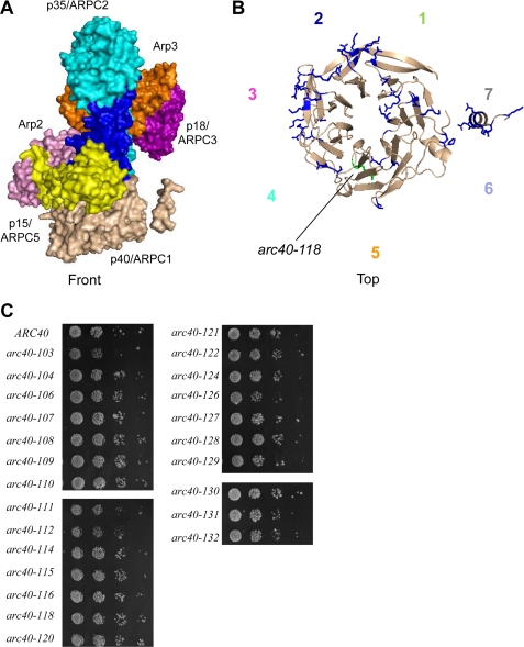
FIGURE 2.
Analysis of initial p40/ARPC1 alanine scan mutations. A, crystal structure of bovine Arp2/3 complex (adapted from Ref. 3), with the seven subunits differently colored: Arp2 (pink), Arp3 (orange), p40/ARPC1 (tan), p35/ARPC2 (cyan), p18/ARPC3 (purple), p19/ARPC4 (blue), and p15/ARPC5 (yellow). B, top view of p40/ARPC1 showing the positions of residues mutated in the initial alanine scan allele collection modeled on bovine ARPC1. The one temperature-sensitive allele (arc40-118; Fig. S1) is green; all other alleles were psuedo-wild type for cell growth and actin organization and are colored blue. The propeller blades are numbered 1–7 counterclockwise in the top view and color-coded to be consistent with Fig. 1. The side chains of residues mutated in arc40 alleles are shown. C, growth of integrated arc40 haploid strains at 25 °C. All of the strains were serially diluted and plated for growth on YPD medium at different temperatures (16, 25, 30, 34, and 37 °C); no defects in growth were observed at any temperature (not shown).

