
Fig. 1. LRRK2 detection by immunoblotting.
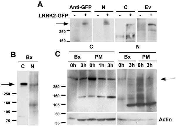
(A) Detection of a LRRK2-GFP reporter gene overexpressed in HEK cells using the three anti-LRRK2 antibodies used in this study. Recognition of the appropriate band is confirmed with an anti-GFP antibody. (B) Detection of the predicted 286 KDa band on a brain homogenate from a temporal lobe biopsy (Bx) by the C- and N-terminal antibodies. (C) LRRK2 detection in human post-mortem samples. Both the C-terminal and the N-terminal antibodies detect the same band of about 286 KDa in human post-mortem brain high salt homogenate extracts from several controls and patients. Since post-mortem (PM) samples from control and diseased brain gave a different pattern to the Bx sample, possibly due to post-mortem degradation, we performed a degradation assay of 1 or 3 hours with portions of the Bx sample and reproduced the same pattern as with the PM samples.