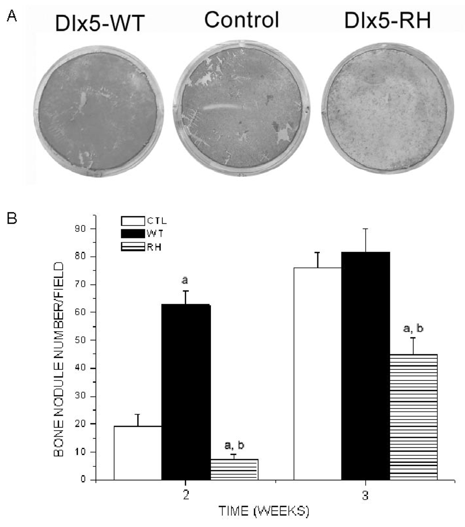
Figure 4.
Effects of RCAS-Dlx5WT, RCAS-Dlx5RH, or empty RCAS vector in bone nodule formation of calvarial cells expressing TVA. Calvarial cells from BSP/TVA mice were infected with RCAS empty vector (Control), wild-type Dlx5 (Dlx5-WT), and mutated Dlx5 (Dlx5-RH). The formation of in vitro mineralization nodules was determined by alizarin red-S histochemical staining, and the number of nodules was counted under a microscope at different time-points (1, 2, and 3 wks). (A) Representative example of alizarin red-S staining 2 wks after infection and osteogenic differentiation. (B) Numbers of nodules obtained 2 and 3 wks after infection and osteogenic differentiation. Results are the mean ± SE from 3 different experiments. Values of p < 0.05 were considered significantly different (ap < 0.05 vs. CTL at every time-point; bp < 0.05 RH vs. WT at every time-point).

