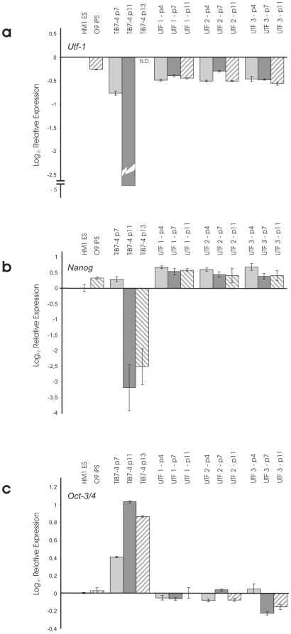
Figure 3. Comparison of relative expressions of Nanog (a), Oct-3/4 (b) and Utf-1 (c) in TiB7-4 wildtype iPS cells at different passages and UTF-Neo selected clones UTF-1, -2 and –3.
Expression levels are normalized to HM-1 ES cells. The Oct-4-Neo selected iPS cell line O9 was included for comparison. N.D.: not detectable.

