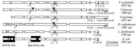
Figure 4.

Schematic alignment of class II reductases with the class I E. coli R1 protein. The alignment was generated with the clustal w version 1.7 (24) program. Single lines show larger gaps introduced in the sequences. The lowest bar gives the length of 120 residues. The arrows show the positions of inteins in the P. furiosus and DR enzymes. The striped area of the DR structure indicates the insertion referred to in the text. The shaded areas show the locations of the two allosteric sites of the E. coli enzyme identified by crystallography (12).
