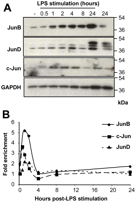
Figure 2. Expression of Jun proteins and jun mRNAs in LPS-stimulated BMDCs.
(A) Expression of the Jun proteins. BMDCs were stimulated by LPS and c-Jun, JunB and JunD levels were assayed by immunoblotting. A representative experiment out of 5 is shown. GAPDH was used as an invariant electrophoresis loading control. JunD classically appeared as 3 bands whose exact molecular natures are still not elucidated. (B) Expression of jun RNAs. BMDCs were stimulated by LPS and c-jun, junb and jund mRNA levels were assayed by qRT-PCR. As inductions of junb and c-jun mRNAs peaked at different times ranging from. 5 to 1.5 hour post-stimulation, depending on the BMDC preparation, no error bar is presented. Instead, 1 representative experiment out of 5 is presented.

