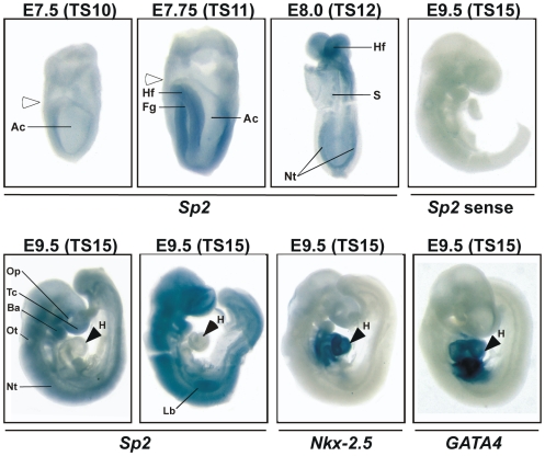
Figure 1. Sp2 mRNA expression in mouse embryos.
Whole-mount in situ hybridization on embryos at the indicated developmental Theiler stages (E7.5-TS10, E7.75-TS11, E8.0-TS12 and E9.5-TS15) with the indicated probes (Sp2, Sp2 sense, Nkx-2.5 and GATA4). The two embryos at E9.5 (TS15) represent two independent in situ hybridization experiments with embryos obtained from different litters. Ac, amniotic cavity; Ba, first branchial arch; Hf, headfold; Fg, foregut; H, heart; Lb, limb bud; Nt, neuronal tissue; Op, optic pit; Ot, otic vesicle; S, somites; Tc, telencephalon. The white arrowheads denote the boundary between embryonic and extraembryonic tissue.

