Figure 2.
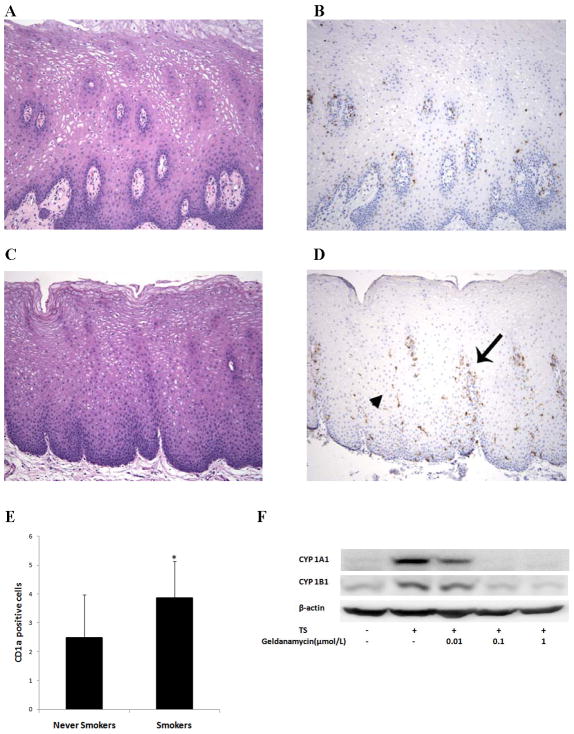
Increased numbers of Langerhans cells were found in the oral mucosa of smokers. Non-neoplastic oral mucosae from never smokers (A) and smokers (C) were morphologically similar, but samples from never smokers showed relatively few Langerhans cells (B) compared to those from smokers (D), which contained numerous Langerhans cells in the peripapillary (arrow) and interpapillary mucosa (arrowhead). [Magnification 100X for panels A–D; panels A and C stained with hematoxylin and eosin; panels B and D stained with CD1a immunostain and hematoxylin]. E, Intraepithelial cells that displayed moderate to strong cytoplasmic staining for CD1a in dendritic-type cellular processes were quantified in the peripapillary, interpapillary and superficial epithelium. A statistically significant increase in the number of CD1a positive cells was found in all three regions in smokers compared to never smokers (P<0.001, <0.001 and =0.032 for peripapillary, interpapillary and superficial areas, respectively). Panel E reflects the total number of CD1a positive cells in the three regions. Columns, means; bars, S.E.; n = 27/group. *, P<0.001. F, MSK-Leuk1 cells were pretreated with vehicle or the indicated concentration of geldanamycin for 2 h. Subsequently, cells received vehicle or TS for 5 h and were then harvested for Western blot analysis. Cellular lysate protein (100 μg/lane) was loaded onto a 10% SDS–polyacrylamide gel, electrophoresed and subsequently transferred onto nitrocellulose. Immunoblots were probed with antibodies specific for CYP1A1, CYP1B1 and β-actin.

