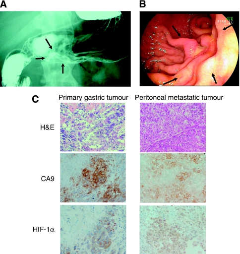
Figure 1.
Upper gastrointestinal series (A) and gastro-fibrscopy (B) showed diffusely infiltrating carcinomas in which ulceration is usually not a marked feature (arrows). Histology of the primary tumour and the peritoneal metastatic tumour showed poorly differentiated adenocarcinoma (C). Primary gastric tumour accompanied by fibrosis. Immunostaining of hypoxia-inducible factor-1α (HIF-1α) and carbonic anhydrase 9 (CA9) showed that hypoxic lesions were present in both the primary tumour and the peritoneal metastatic tumour (C).

