
Figure 6.
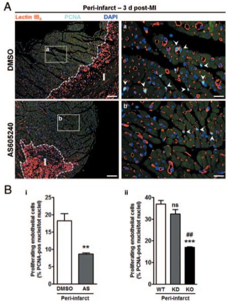
Targeting of PI3Kγ inhibits EC proliferation in infarcted hearts. Representative immunofluorescence images (A) show the fraction of proliferating ECs in PI zone of DMSO- or AS-treated hearts (a and b, respectively) 3 days post-MI. Proliferating ECs (arrowheads) are identified by lectin IB4 (red) and positivity for proliferating-cell nuclear antigen (PCNA) (light blue). Nuclei are stained by DAPI (blue). Scale bars: left, 100 μm; right, 20 μm. I indicates infarct; unspecific lectin IB4 binding delimiting infarcted area. B, Bar graph illustrates the number of proliferating ECs in PI zone of DMSO- or AS-treated mice (i) or of WT, KD and KO mice (ii). n=4 to 6 mice in each group. **P<0.01 vs DMSO; ***P<0.001 vs WT; ##P<0.01 vs KD; ns, not significant vs WT.