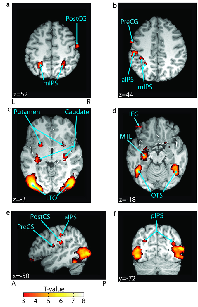
Figure 3.
Cross-modal integration. Regions significant in a block-level contrast of AVs > AVo (FDR p < .05, n = 25) are overlaid on a representative brain. Significant regions include (A) the right postcentral gyrus (PostCG) and the bilateral middle intraparietal sulcus (mIPS); (B) the precentral gyrus (PreCG) and the anterior/ middle IPS (aIPS/mIPS); (C) the bilateral putamen, the tail of the caudate nucleus (caudate), and the lateral temporal–occipital boundary (LTO); (D) the left inferior frontal gyrus (IFG), the left medial temporal lobe (MTL), and the bilateral occipito-temporal sulcus (OTS); (E) the left precentral sulcus (PreCS) and the left postcentral sulcus (PostCS); and finally, (F) the bilateral posterior IPS (pIPS). MNI coordinates are reported in white text in each panel. A = anterior; P = posterior; L = left; R = right.

