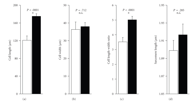
Figure 3.
Geometrical Dimensions of Human Ventricular Myocytes from Failing and Non-Failing Hearts. Cell length and width were measured from transmitted light images of each cell taken with a 60× lens at 1- to 2-fold zoom (a,b). Cell length-width ratio was calculated from these data (c). Sarcomere length was measured from 6–9 sarcomeres in the transmitted light images (d). Open bars show the dimensions for myocytes from non-failing hearts (11 cells, 2 hearts), closed bars represent cells from failing human hearts (37 cells, 4 hearts). Calculated p-values as given, stars symbolize statistical significance.

