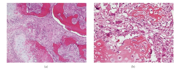
Figure 3.
(a) Low-power photomicrograph of the primary tumor, showing irregularly arranged woven bone trabeculae and atypical cartilage islands along with intervening fibrous tissue. (b) High-power photomicrograph, showing irregular osteoid seams with atypical osteoblasts and atypical chondrocytes within the cartilage matrix. These findings are consistent with low-grade osteosarcoma.

