
FIGURE 4.
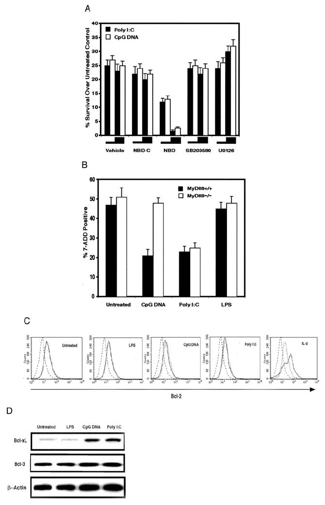
Poly(I:C) or CpG DNA-mediated survival requires NF-κB activation and is associated with Bcl-xL up-regulation but only CpG DNA-mediated survival is MyD88 dependent. A, Purified DO11.10 CD4+ T cells, first activated with pOVA-pulsed APCs for 16 h and then purified by magnetic beads, were treated with poly(I:C) (90 μg/ml), CpG DNA (30 μM), or left untreated and coincubated with NBD (5 or 20 μM), NBD-C (5 or 20 μM), SB203580 (2.5 or 10 μM), U0126 (2.5 or 10 μM), or vehicle control for 48 h. Viability is expressed as the percentage difference of 7-AAD excluding cells between TLR ligand-treated and untreated cells. Results are from four independently performed experiments ± SEM. B, Magnetic bead-purified CD4+ T cells from MyD88−/− and wild-type control littermates (MyD88+/+) were activated with plate-bound anti-CD3 plus anti-CD28 mAbs, washed, and replated with either poly(I:C) (30 μg/ml), CpG DNA (10 μM), LPS (100 ng/ml), or left untreated for 72 h. Death was assessed as the percentage of 7-AAD positively stained cells. Results are from three independently performed experiments ± SEM. C, Purified activated DO11.10 CD4+ T cells, prepared as in A, were treated with either poly(I:C) (90 μg/ml), CpG DNA (30 μM), LPS(1 μg/ml), PGN (30 μg/ml), IL-2 (50 U/ml), or left untreated for 48 h. TLR ligand-treated cells (solid lines), IL-2-treated cells (solid line), or untreated cells (dotted line) were stained for Bcl-2 or an isotype control (dashed line). The result is representative of three independently performed experiments. D, Purified activated DO11.10 CD4+ T cells, prepared as in A, were treated with either poly(I:C) (90 μg/ml), CpG DNA (30 μM), LPS (1 μg/ml), or left untreated 48 h, lysed, and analyzed by Western blot with Abs specific for mouse Bcl-xL, Bcl-3, or β-actin.