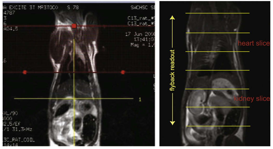
Fig. 4.
T2 weighted Sagittal FSE images from one of the rat studied. The spectral-spatial band is placed across the upper torso of the animal to saturate the 13C metabolite signal from the heart and lung, and the placement is performed using the interactive graphic Rx (left). The spatial localization in the dynamic MRS experiment achieved by the echo-planar readout in relation to the anatomy is also shown (right).

