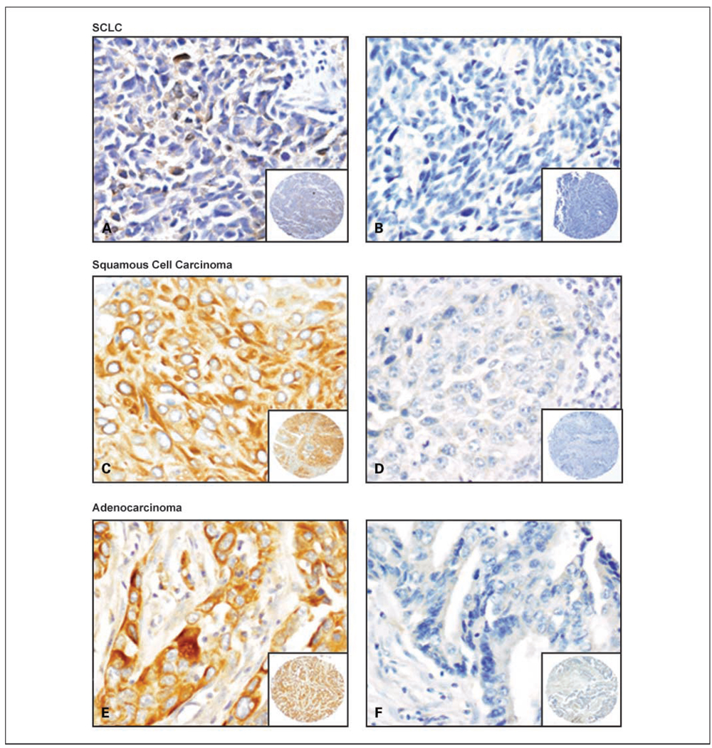
Fig. 1.
Representative examples of Fus1 immunohistochemical staining of lung cancer specimens. SCLC with reduced (A) and negative (B) Fus1 expression. Squamous cell carcinoma with high (C) and negative (D) Fus1 expression. Adenocarcinoma with high (E) and negative (F) Fus1 expression. A to F, original magnification, ×400 (pictures), and ×40 (insets).

