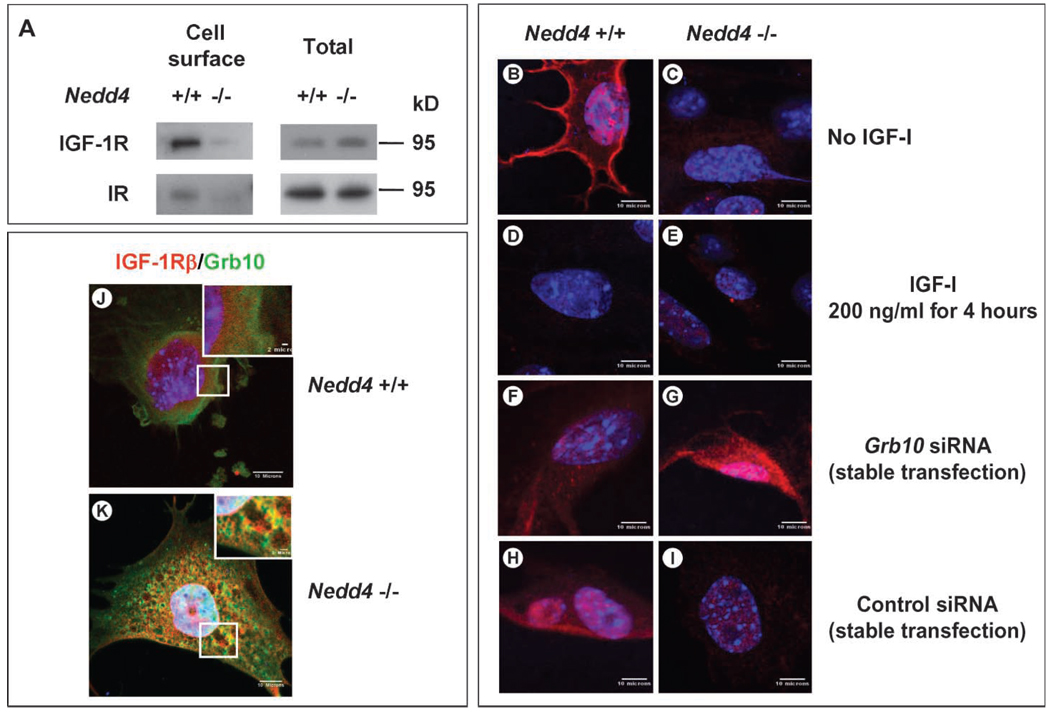
Fig. 7.
Cell surface IGF-1R concentrations change when Nedd4 and Grb10 protein expression are altered. (A) Decreased cell surface IGF-1R and IR in Nedd4-deficient MEFs. Cell surface proteins were isolated by biotin labeling and affinity purification and detected by immunoblotting. (B to I) IGF-1R concentrations at the plasma membranes of Nedd4+/+ and Nedd4−/− MEFs were determined in the absence or presence of an siRNA against Grb10 (rabbit anti–IGF-1Rα antibody was used at 1:100, Santa Cruz Biotechnology). (B and C) No IGF-1 stimulation. (D and E) Cells stimulated with recombinant IGF-1 (at 200 ng/ml, Sigma) for 4 hours. (F and G) Cells stably transfected with siRNA targeting Grb10. (H and I) Cells stably transfected with control siRNA. IGF-1R was not detectable on Nedd4−/− cells, but Grb10 knockdown restored IGF-1R to the plasma membrane. (J and K) Grb10 and IGF-1R are partially colocalized in Nedd4−/− MEF cells. MEFs grown in DMEM + 10% fetal bovine serum were fixed with 2% paraformaldehyde for 30 min at room temperature. Grb10 (green) and IGF-1R (red) were detected with monoclonal antibody against Grb10 and rabbit polyclonal antibody against IGF-1Rβ (Santa Cruz Biotechnology). Scale bar, 10 µm in the main figures and 2 µm in the insets.

