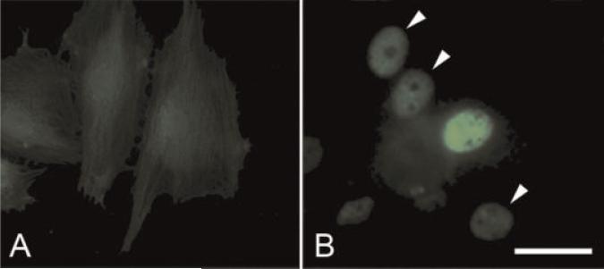
Fig. 6.
Immunofluorescent labeling for obscurin-associated protein kinase in the permanent line of H9c2 cells transfected with a non-coding control construct (A) and transfected with His-tagged construct encoding obscurin-associated kinase (B). Note intense fluorescent labeling of anti-His n the nucleus of a transfected cell in the center of the field in panel B. Arrowheads in B show the location of weakly and moderately positive nuclei. Bar, 30 μm. [Color figure can be viewed in the online issue, which is available at www.interscience.wiley.com.]

