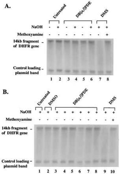
Figure 2.

Alkaline-agarose gel electrophoresis of DB[a,l]PDE-reacted B11 cell DNA and DNA from DB[a,l]PDE-treated B11 cells. CHO-B11 DNA and B11 cells in culture were treated with DB[a,l]PDE. The DNA was incubated in the presence or absence of sodium hydroxide (indicated above the lanes) for 30 min at 37°C (see Materials and Methods). DMS-methylated DNA was included as a positive control for the detection of AP sites. (A) KpnI-restricted DNA: untreated (lanes 1 and 2); reacted with 76 μM (+)-syn-DB[a,l]PDE (lanes 3 and 4); reacted with 13 μM (−)-anti-DB[a,l]PDE (lanes 5 and 6); reacted with 0.1 M DMS (lane 7 without methoxyamine treatment and lane 8 with methoxyamine treatment). (B) KpnI-restricted DNA from CHO-B11 cell cultures treated with: no treatment (lane 1); DMSO (lane 2); 1.4, 4.3, or 8.5 μM (+)-syn-DB[a,l]PDE (lanes 3–5); 1.4, 4.3, or 8.5 μM (−)-anti-DB[a,l]PDE (lanes 6–8); or DMS [without methoxyamine (lane 9) or with methoxyamine (lane 10)]. All samples underwent electrophoresis on a 0.5% alkaline-agarose gel and were probed with a 32P riboprobe for the transcribed strand of the 14-kb fragment of the DHFR gene.
