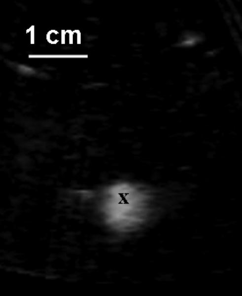
Fig. 2.

Prior to the treatment, a bubble cloud was generated in a water bath, which was shown as a hyperechoic zone on an ultrasound image and used for target localization. Ultrasound was delivered from the top to the bottom. The front center of the hyperechoic zone was marked as the focal zone (‘x’). The image was collected using a 5 MHz imaging probe inserted in the central hole of the therapeutic transducer.
