
Fig. 4.
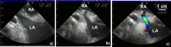
a) Before treatment ultrasound image of the intact atrial septum (marked by arrow). b) Representative ultrasound image of the ASD generated by histotripsy. The ASD appears as a dark channel through the atrial septum (marked by arrow). c) The Doppler color flow mapping showing a blood jet through the ASD. All images were collected using a 10 MHz imaging probe.