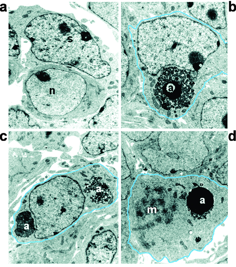
Figure 2. Electron micrographs of apoptotic bodies engulfed and ingested by SGC precursors in embryonic E12.5 DRGs.
(a) A healthy neuron [n] characterized by finely dispersed chromatin and abundant cytoplasm adjacent to a SGC precursor [s] with a characteristic pleomorphic nucleus with chromatin clumps. (b) An apoptotic cell [a] engulfed by a SGC precursor. (c) Debris of two ingested cells [a] inside a SGC precursor. (d) An ingested apoptotic cell present in a cell that is undergoing mitosis (m; mitotic figure, condensing chromosomes).

