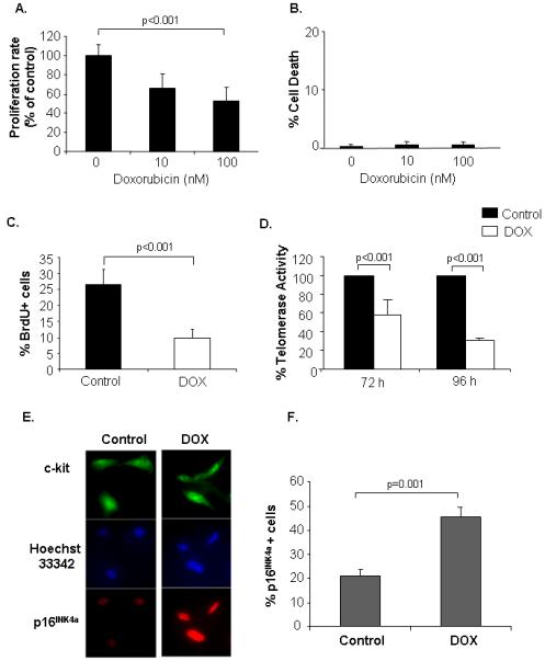
Figure 8.

Doxorubicin treatment inhibits proliferation of isolated CPCs. Cells were treated with doxorubicin for 72 h and then assessed for (A) cell proliferation by MTT assay (n=3), and (B) cell death by Trypan Blue exclusion assay (n=3). C. Cells were treated with 100 nM doxorubicin and proliferation was determined by BrdU incorporation (n=4). D. Cells were exposed to vehicle or 100 nM DOX for 72 h or 96 h and then assayed for telomerase activity using the TRAP assay (n=3). E. Isolated CPCs were treated with 100 nM DOX for 72 h and then fixed and stained for the presence of c-kit and p16INK4a. F. Quantitation of p16INK4a positive cells (n=4).
