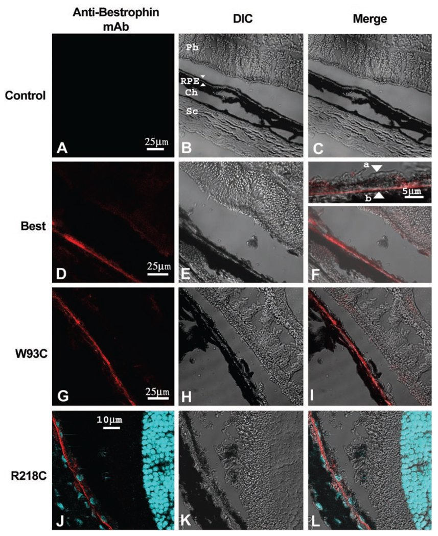
FIGURE 1.
Normal localization of bestrophin and bestrophin mutants. The localization of bestrophin (D–F) or bestrophin mutants (W93C, G–I; R218C, J–L) was determined by immunofluorescence staining and confocal microscopy in eyes injected with a control (Null, A–C) vector or with adenovirus vectors encoding bestrophin (WT), or bestrophin mutants (W93C, R218C). Cryosections of rat eyes preserved in cold methanol were stained with monoclonal antibody E6-6 and a Texas-red–conjugated secondary antibody (red). For eyes expressing bestrophin-R218C, nuclei were stained with DAPI (blue). Confocal images of sections stained for bestrophin (Mab, A, D, G, J, red staining) and corresponding differential interference contrast (DIC, B, E, H, K) images are shown. When images were merged (Merge, C, F, I, L), bestrophin staining exhibited a pattern consistent with localization to the RPE (B, arrows). Inspection at higher magnification (F, inset) demonstrates that the protein was predominantly in the basolateral plasma membrane of the RPE (F, inset, apical membrane, a; basal membrane, b). Other cell types in the sclera (Sc), choroid (Ch), or neurosensory retina (Ph) did not express bestrophin. Also note that the antibody was specific for the human form of bestrophin and did not react with the endogenous protein in the uninjected eye.

