Figure 8.
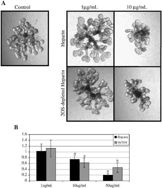
Heparin and 2OS-depleted heparin titration curve on isolated ureteric bud (UB) branching morphogenesis. A: Phase contrast micrographs of UBs isolated from E13 kidney rudiments, suspended in a 3-dimensional extracellular matrix and cultured in the presence of BSN conditioned media and 125ng/ml GDNF and 125ng/ml FGF1 and varying concentrations of heparin and 2OS-depleted heparin. Images at 10×. B: Graphical analysis of the average number of UB tips as a percentage of control. At a concentration of 10μg/mL and higher, both heparin and 2OS-depleted heparin significantly inhibit UB branching indicating that the binding of factors involved in UB branching are not markedly affected by the 2-O sulfate modification. Mean ± SD, n>3, *p<0.05 compared to control.

