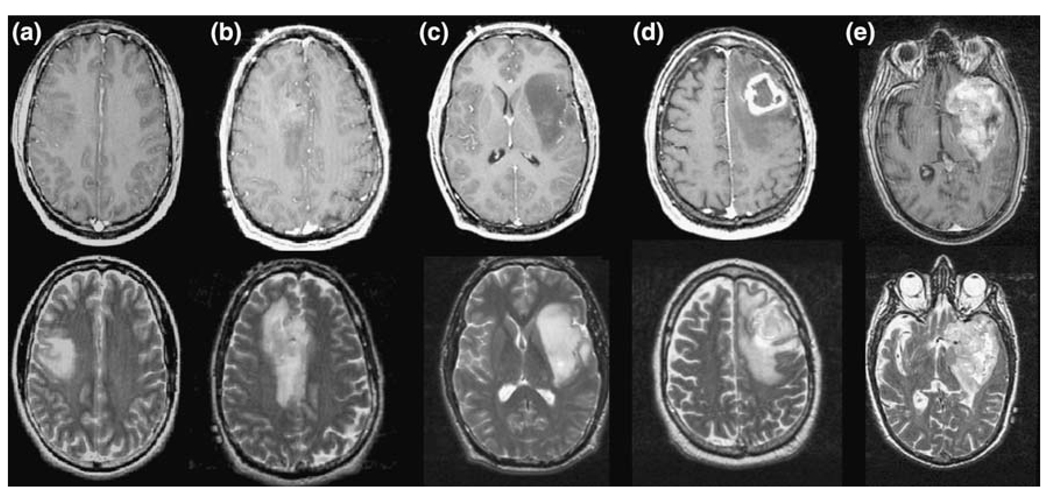
Fig. 1.
T1-weighted post-Gadolinium and T2-weighted images of Grade II (a, b), III (c) and IV (d, e) glioma. The volumes of the overall T2 hyperintensity are large in all three cases, but the T1-weighted images shows larger contrast enhancement and/or hypointense necrotic regions for the grade IV glioma, no enhancement for the grade II lesion and marginal or no enhancement for the grade III gliomas

