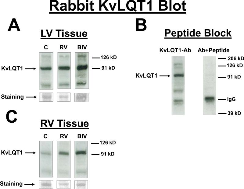
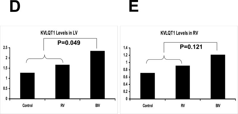
Figure 4.
KvLQT1 protein blot from the LV (A) and the RV (C) tissues for the 3 rabbit study groups (C, RV, and BIV). KVLQT1 bands as well as coomessie-stained bands used to normalize for the protein load are shown. Also the KVLQT1 bands for the 3 study groups are from the same blot with identical exposure. B: KVLQT1-peptide block, where the blot was exposed to anti- KVLQT1+petide (1:3 ratio). Panels D and E are the graphic representation of KVLQT1 protein levels from LV (D) and RV (E) tissues for the 3 study groups. Note the statistically significant increase in KVLQT1 levels in the BIV group compared to the combined RV paced and control groups in the LV but not RV extracted tissue.


