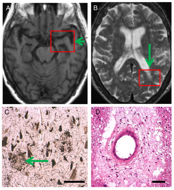
Fig. 6.
Diabetes Case 4–82-year old female diabetic patient with dementia and Alzheimer’s disease diagnosis during life (last MMSE score=19). A and B show the final MRI that was obtained 4 years prior to the patient’s demise. This already showed hippocampal atrophy (arrow in A) but also some periventricular white matter lesions (such as in arrow in B). Photomicrographs show the histopathological features from the red boxes. C depicts a section from CA1 field of the hippocampus stained with the Gallyas silver impregnation technique and shows severe involvement by Alzheimer’s-type neuritic plaques (arrow) and many NFTs. This patient had Braak stage 6 and satisfied CERAD criteria for “Definite Alzheimer’s disease” by pathology. In addition to the AD pathology, there was also some cerebrovascular disease including areas with rarefaction of white matter. D is a section from the left parietal lobe (box in B) which shows an expanded Virchow–Robin space with organized cellular and acellular material. In an 82-year old patient such as this, some degree of concomitant pathology is the rule and not the exception. Scale bars=150 μm in C, 100 μm in D.

