
Fig. 8.
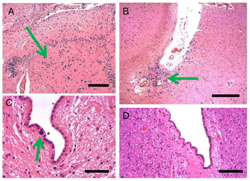
Diabetes Case 6–72-year old female patient with poorly controlled diabetes (two confirmed readings in excess of 150 mg/dl) and mild cognitive decline. In this case, the hippocampal formation showed many corpora amylacea including in the cornu ammonis subfields. These are shown in the CA4/dentate gyrus area, where there is some effacement of the normal cytoarchitecture in association with the presence of many corpora amylacea. B shows the crux of the inferior horn of the lateral ventricle, which also contained many corpora amylacea. By contrast, in Control Cases 1 and 2, there were few (arrow in C) or no corpora amylacea there. Scale bars: 300 μm in A and B, 150 μm in C and D.