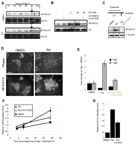
Fig. 2.
Initial exposure to RA induces Fgf8/Erk signalling during ES cell differentiation. (A) On day 1 of differentiation RA addition causes Erk activation by 8 hours, western blots represent two independent experiments, arrows indicate elevated dP-Erk in RA but not DMSO conditions. (B) Treatment with 0.5 mM of each of LG100815 and LG101208 blocks the ability of 50 nM RA to induce an increase in dP-Erk levels by 8 hours on day 1 of differentiation. This experiment was performed twice with the same result. (C) PD173074 blocks RA induction of dP-Erk by 24 hours on day 1 of differentiation. This experiment was performed twice with the same result. α-Tubulin was used as a loading control. (D) RA-treatment on day 1 increases dP-Erk in differentiating cells. Scale bar: 50 μm. (E) On day 1, RA reduces Fgf4 but increases Fgf8, and these effects are both inhibited by RAR/RXR antagonists. Results are weighted means ±s.e.m. from two independent experiments performed in triplicate. (F) Fgf8 transcripts rise in response to RA during day 1 even when differentiation is blocked by PD173074, representative of two experiments, ±s.e.m. (G) The ability of RA to induce dP-Erk1/2 is attenuated with 2.5 μg/ml anti-Fgf8 blocking antibody for 24 hours. dP-Erk was quantified by fluorescence immunoblotting. This experiment was carried out twice; result shown is the mean from a representative experiment performed in triplicate ±s.e.m.

