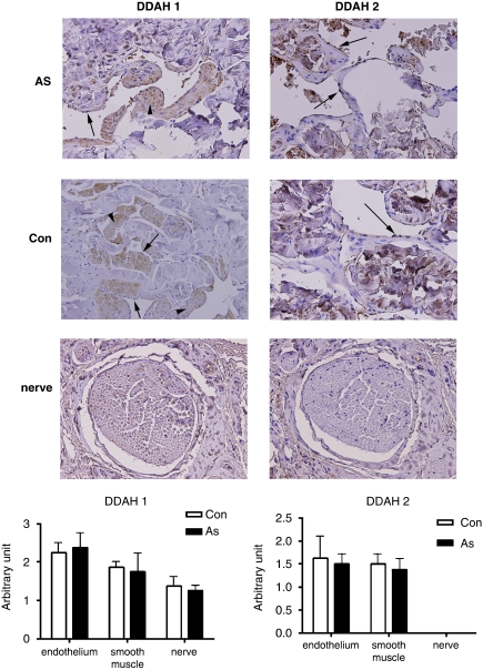
Figure 3.
Immunohistochemical evaluation of both dimethylarginine dimethylaminohydrolase (DDAH) isoforms in rat cavernosal tissues. Immunoreactivities are seen in brown. Upper two rows revealed representative pictures from normal control (Con) and atherosclerosis (AS) groups. Compared to DDAH 2, which showed only modest endothelial staining (arrow), DDAH 1 showed a greater staining intensity and was found in both endothelial (arrow) and trabecular smooth muscle (arrowhead). Representative neural picture in third row was from Con group. Similar findings were observed in AS group, so only pictures from Con group were displayed. Abundant staining for DDAH 1 was identified in neural cells, whereas no immunostaining was observed for DDAH 2. Comparison of staining scores showed similar staining intensity of both isoforms in the Con and AS groups, as shown in densitometry.

