Figure 4.
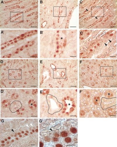
Synchronous cell division in Pkd1 and Pkhd1 mutant mice. (A through F′) Longitudinal (A through C and A′ through C′) and transverse (D through F and D′ through F′) sections from kidneys of newborn mice stained with PCNA. Contiguous stretches of PCNA-positive nuclei suggestive of synchronous cell divisions are present in WT (A, A′, D, and D′) and Pkd1flox/−;Ksp-Cre (B, B'′, E, and E′) kidneys. Pkhd1del4/del4 kidneys (C, C′, F, and F′) show a more interrupted and discontinuous pattern of PCNA-positive nuclei in elongating tubules (C and C′, arrowheads). Tubules in transverse section were often uniformly negative (outlined) or uniformly positive (*) for PCNA in WT and Pkd1flox/−;Ksp-Cre kidneys (D, D′, E, and E′) but not in Pkhd1del4/del4 kidneys (F and F′, *). Dilating tubules of early cysts in Pkd1flox/−;Ksp-Cre kidneys (B′ and E′, *) continue to show a synchronous pattern of PCNA expression. A′ through F′ are digitally magnified views of the respective boxed regions in A through F. PCNA incorporation in Pkhd1del4/del4 kidneys occasionally showed misoriented cell (G and G′, black arrowhead); white arrowheads show discontinuous pattern of PCNA staining. Bars = 50 μm in A through F, 15 μm in A′ through F′, and 5 μm in G and G′.

