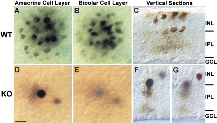
Figure 5. Gap Junctions between AIIs and AII and On CBs Are Disrupted.
(A and B) In WT mouse retina, neurobiotin (mw = 284 Da) injected into individual AII amacrine cells is detectable both in adjacent AIIs (A) and in the overlying CBs (B), consistent with the notion that gap junctions couple these cell types.
(D and E) In dramatic contrast, neurobiotin is completely restricted to the injected AII in the Cx36 KO.
(C, F, and G) Vertical sections were used to confirm the identities of injected cells.
Scale bars equal 10 μm.

