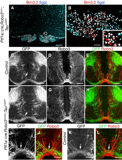
Figure 8. Analysis of Ptf1a::cre;Robo3lox/lox;TaumGFP mice.
(A and B) Coronal sections of the hindbrain at the level of inferior olive of E16 Ptf1a::cre;TaumGFP embryo labeled with anti-βgal and Brn3.2 antibodies. Some Brn3.2-positive neurons in the inferior olive do not express the βgal (arrowheads and inset). (C–L) Coronal sections of the spinal cord at the level of the forelimbs in E13 Ptf1a::cre;Robo3lox/+;TaumGFP (C–E, K, and L) or Ptf1a::cre;Robo3lox/lox;TaumGFP (F–H, I, and J) embryos labeled with anti-GFP and anti-Robo3 antibodies. Most GFP-positive axons are in the dorsal spinal cord, and only a small subset of GFP-positive axons (short arrows) cross the floor plate in Ptf1a::cre;Robo3lox/+;TaumGFP (C and K) but do not express Robo3 (E and L). This subset of GFP-positive commissural axons is still observed in Ptf1a::cre;Robo3lox/lox;TaumGFP embryos (F and J). Scale bars represent 100 µm except in (B and I–L), where they indicate 50 µm.

