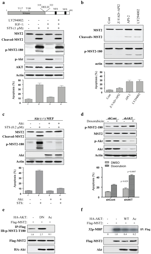
Figure 1. IGF1/PI3K/Akt inhibits MST2 cleavage, activation and MST2-induced cell death.
(a) IGF1 inhibits MST2 through the PI3K/Akt pathway. Top panel is domain structure of MST2. DELD is a caspase cleavage motif and NES stands for nuclear export signal. Middle panels are immunoblots of COS7 cells, which were treated with IGF1 (50 µM) or/and LY294002 (20 µM) for 30 min prior to exposure to STS (1 µM) for 1 h, with indicated antibodies. Bottom panel shows the apoptosis measured with Tunel assay in three experiments in triplicate. (b) Inhibitors of PI3K (LY294002) and Akt (API-2) activate MST2. After treatment with indicated compounds for 3 h, COS7 cells were subjected to immunoblotting with indicated antibodies (upper panels) and apoptosis analysis (bottom). (c) Reconstitution of Akt in Akt1-null MEFs reduces MST2 activation induced by STS. Akt1-knockout MEFs were infected with adenovirus expressing Akt and then treated with 0.2 µM STS for 1h. Immunoblotting (upper) and apoptosis (bottom) analyses were performed as described above. (d) Knockdown of Akt enhances doxorubicin-activated MST2. MDA-MB-468 cells were transfected/treated with Akt/shRNA together with or without doxorubicin, and then subjected to immunoblotting (upper) and Tunel (bottom) analyses. (e) and (f) Constitutively active (Ac) Akt inhibits but dominant-negative (DN) Akt induces MST2 activation. COS7 cells were transfected with indicated plasmids. Following 48 h of incubation, cells were assayed for autophospho-T180 (top panel of e) and in vitro MST2 kinase using MBP as substrate (top panel of f). Middle and bottom panels show expression of the transfected plasmids.

