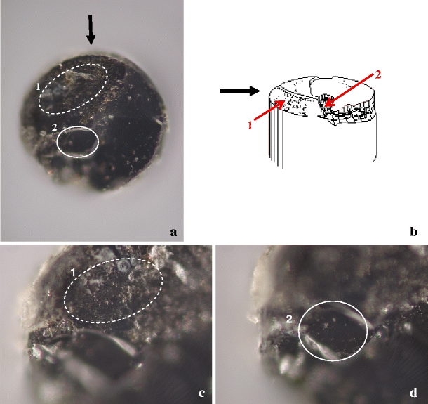
Fig. 5.

Tip of EVLA fibre 20 used at 1,470 nm photographed through a microscope; the fibre diameter is 0.6 mm. a Overview ( × 10). Two damaged areas (1 and 2) are indicated. b Artist’s impression. Black arrow indicates the direction of view indicated in a. The red arrows point to areas 1 and 2. c Image focused on irregular and cracked area 1 (×20). d Image focused 29 μm lower than c on area 2 (×20)
