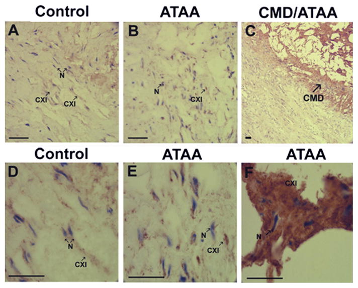
Fig 2.

Immunohistological analysis of collagen α1(XI). (A) Control tissue and (B) ascending thoracic aortic aneurysms (ATAA) tissue, at ×200 original magnification. (C) Cystic medial degenerative (CMD) lesion at ×50 original magnification. (D) Control tissue, (E) ATAA tissue, and (F) CMD lesion from ATAA at ×630 original magnification. Tissue sections stained with antibody directed to collagen α1(XI) demonstrated staining within the tunica media in both control tissues and ATAA. Presence of collagen α1(XI) is indicated by brown staining (indicated by arrow CXI). Tissues were counterstained briefly with hematoxylin to stain nuclei (blue, indicated by arrow N). An increase in staining for collagen α1(XI) is shown within regions of CMD lesions. Scale bars in A to F are 100 μm.
