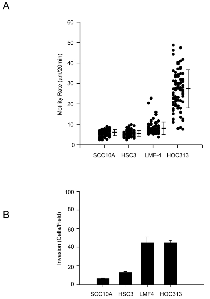
Figure 1.
Relative motility and invasion of oral SCC cells. (A) For cell motility assays, head and neck SCC cell lines were plated on 1 µg/ml collagen I substrates. Time-lapse images were taken at 20 min intervals for 3 hr. Cell motility rates were analyzed from at least 60 cells. The data represents the mean and S.D of cell motility rate for each cell type (B) Head and neck SCC cell lines were assessed for the invasion of collagen I gels as described under the “Material and Methods”. The data represent the mean and S.D. of cells that invaded through the collagen gel barrier.

