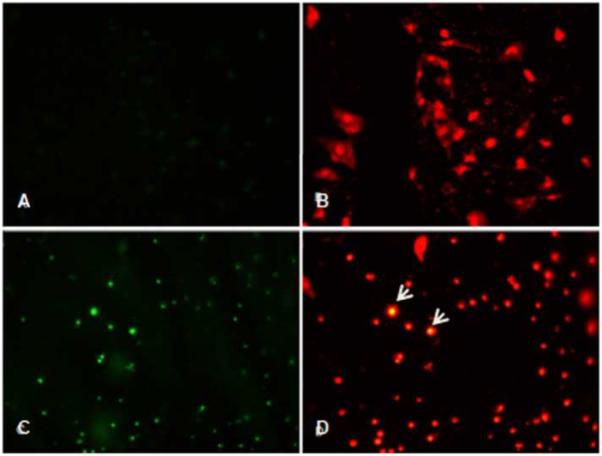
Figure 4.

EGCG-mediated apoptosis assessed by TUNEL staining. Apoptotic nuclei are detected with green fluorescence and all nuclei stained with propidium iodide (red fluorescence) in ELT3 cells at 0μM (A, B) and 100μM(C, D) EGCG. Arrows indicate prominent apoptotic nuclei that are double stained. Magnification 200×
